

What are the stages of diabetic retinopathy?
There are four. The first three are called nonproliferative diabetic retinopathy: mild, moderate, and severe. The fourth is proliferative diabetic retinopathy, when new blood vessels start to grow on the retina. Diabetic macular edema, a swelling in the center of the retina, can show up at any stage.
When we do find diabetic retinopathy, finding it in the early stages is the good news, because there is time to act and to prevent vision loss.
| What is happening in the retina | What you may notice | What we do |
|---|---|---|
| Mild nonproliferative | ||
| Tiny bulges form in the small blood vessels and may leak a little. | Usually nothing. | Photograph the retina, keep the picture, and see you again in a year unless we say sooner. |
| Moderate nonproliferative | ||
| More vessels swell and leak, and some become blocked. | Usually nothing, unless the macula swells. | See you back sooner, scan the macula with OCT, and send a report to your doctor. |
| Severe nonproliferative | ||
| Many vessels are blocked, and parts of the retina lose their blood supply. | Often still nothing. | Bring in a retina specialist and keep a close watch between visits. |
| Proliferative | ||
| Fragile new blood vessels grow. They can bleed, and scar tissue can form. | Floaters, dark spots, blurry vision, or sudden vision loss. | Refer you promptly to a retina specialist for laser treatment or injections. |
| Diabetic macular edema (any stage) | ||
| Fluid leaks into the macula, the part of the retina you read with. | Blurry or wavy central vision, or faded colors. | Measure the swelling with OCT and refer you for treatment when it needs it. |
The stages of diabetic retinopathy, and what we do at each one
What is diabetic retinopathy, and what causes it?
Diabetic retinopathy is damage to the tiny blood vessels in the retina, the thin layer at the back of the eye that turns light into the picture your brain sees. Years of high blood sugar weaken the walls of those vessels. Weak vessels leak fluid and blood, and some close off.
When part of the retina loses its blood supply, it sends out signals to grow new blood vessels. That sounds helpful, but these abnormal blood vessels are fragile and bleed easily. That is the line between nonproliferative diabetic retinopathy and proliferative diabetic retinopathy.
In proliferative diabetic retinopathy, the new blood vessels can bleed into the clear gel that fills the eye. That is called a vitreous hemorrhage, and it shows up as sudden floaters or a dark haze. Scar tissue from the new vessels can also pull on the retina and, in advanced diabetic retinopathy, lead to a retinal detachment.
It can happen with type 1 or type 2 diabetes. The longer you have had diabetes, the higher the risk. Blood sugar that runs high, high blood pressure, and high cholesterol add to it. Smoking adds too.
What are the symptoms of diabetic retinopathy?
In the early stages, none. Most people with diabetes who have early retinopathy do not notice anything. Your vision can feel perfectly fine while the blood vessels in your retina are changing. That is why your primary care doctor keeps asking for a yearly eye exam, even when you see well.
Symptoms tend to come later, often with macular edema or the proliferative stage. People describe blurry vision that comes and goes, new floaters, dark or empty areas in their sight, colors that look faded, or trouble reading fine print. Blurry vision with diabetes is not always retinopathy, though. A blood sugar swing can blur your glasses prescription for a while, and the exam tells us which one you have.
Our own appointment book from September 2025 through August 2026 shows about three visits in ten with us are medical visits, not glasses checks. Office visits, scans, and follow-ups like these are a normal part of our week, so you are in practiced hands.
What is diabetic macular edema?
Macular edema is swelling in the macula, the small center of the retina that gives you sharp vision for reading and faces. In diabetic macular edema, damaged blood vessels leak fluid into it. It is the most common reason diabetes blurs central vision.
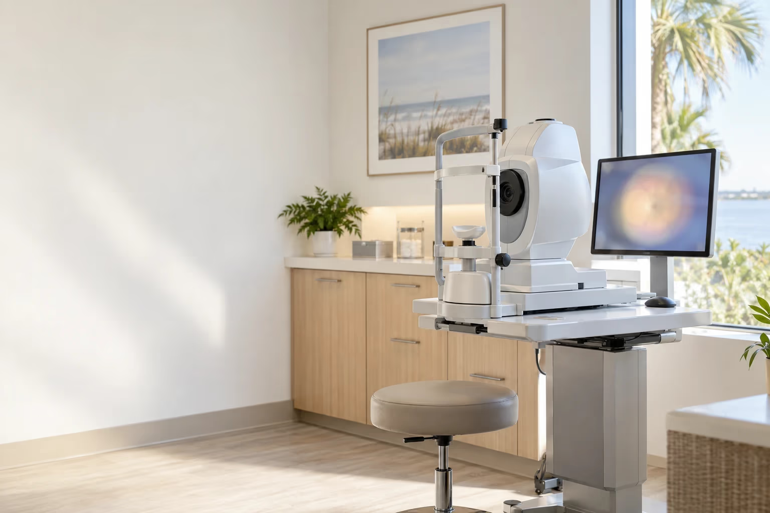
You can have retinopathy without macular edema, and macular edema can start before the retinopathy looks severe. We check for it at every diabetic eye exam. The OptoVue OCT scanner reads the layers of the retina in under a second and shows us how thick the macula is, so we can see swelling before it blurs your reading.
It is not the same as macular degeneration, which is an age-related condition. If you want to know more about that one, we treat macular degeneration too.
What happens at a diabetic eye exam in Titusville?
A diabetic eye exam is a full eye exam with your pupils dilated, so we can see the whole retina out to its edges. We dilate patients with diabetes once a year, because early changes often start at those edges.
- 1
Dilating drops
The drops open your pupils. They take a little while to work, so plan on about an hour with us.
- 2
A wide picture of the retina
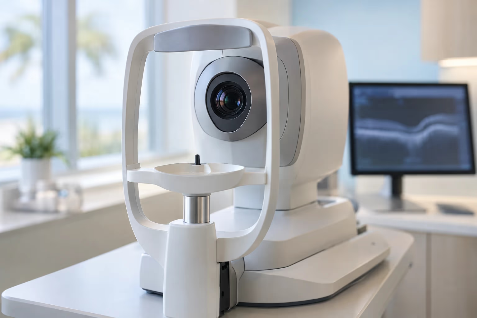
The Optos Optomap takes a wide photo of your retina. We show it to you on the screen and keep it to compare next year.
- 3
A scan of the macula
When we need to measure swelling, the OCT scan shows the retina layer by layer.
- 4
Pressure and the optic nerve
Diabetes raises the odds of glaucoma, so we check your eye pressure and the optic nerve in the same visit.
- 5
Your results, explained
We tell you what we found and which stage, if any, and when we want you back. We can send a report to your primary care doctor.
Your near vision stays blurry and bright light bothers you for a few hours afterward, so bring sunglasses. Here is how long eye dilation lasts if you are planning the drive home.
If we ask to see you back for a scan, book it before you leave. Testing visits ran about a sixth above a usual month in May, June, and August, so summer times fill first.

Can diabetic retinopathy be reversed?
Damage that has already happened usually cannot be undone. Retinopathy can be slowed, though, and treatment can stop leaking and bleeding before they take more vision. Macular swelling often goes down with treatment. Early treatment gives you the most options, which is why the yearly exam matters.
In the mild and moderate stages, the work is mostly yours and your primary care doctor's: managing your diabetes and keeping your blood sugar, blood pressure, and cholesterol where your doctor wants them. That slows diabetic retinopathy more than anything we can do in the exam chair. We watch the retina and send the findings along.
When retinopathy reaches a stage that needs treatment, we refer you to a retina specialist. Treatment is usually injections of medicine that calm leaky vessels, often called anti-VEGF, or laser treatment to seal them. Bleeding that does not clear may need surgery called a vitrectomy. We do not do those procedures here. We send your images along and keep up your yearly exams and everything else your eyes need.
Does Medicare cover a diabetic eye exam in Brevard County?
Usually, yes. Because you have diabetes, the exam is a medical visit. It is billed to Medicare or your medical insurance, not your vision plan, and your usual copay applies. Between October 2025 and May 2026, about one exam in seven we did was billed to Medicare or a Medicare Advantage plan, so we know that paperwork well.
We also take medical plans such as Humana, Blue Cross Blue Shield, Aetna, Cigna, UnitedHealthcare, and Tricare. If you want new glasses at the same visit, the prescription part may go to a vision plan like VSP or EyeMed, or be paid separately. Call us with your plan name and we will check your benefits first. Our insurance page lists every plan we work with, and eye exams after 65 explains the routine and medical split.
Where can I get a diabetic eye exam in Titusville?
At either of our offices, and all four of our doctors do them. Dr. Greg Aker and Dr. Amanda Aker see patients at Aker Eye Downtown, 338 S Washington Ave, on the second floor with an elevator. Dr. Sarah Koehnemann and Dr. Jennifer Bradley see patients at Aker Eye Playalinda, 1114 S Washington Ave, all on one floor. The Optomap and the OCT are part of the exam at both.
November is Diabetes Awareness Month, and it has been the quietest month on our schedule all year. If your diabetic eye exam is due, this is an easy month to get a time. Call us at (321) 269-2021 or book online, and bring the name of the doctor who wants the report.